A Quick Tour
through the Brain
Begin
Small: Neurons
The
brain is made up of neurons. Neurons are the functional units in the brain and
throughout the nervous system. There are more than 180 billion neurons with at
least 80 billion involved in cognitive processes. Each neuron connects with
hundreds of other neurons, and so we have vast potentials for an enormous
amount of interactions occurring simultaneously.
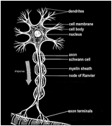
Neurons
have two main functions: They process certain chemicals within them, and they
communicate with other neurons. This communication process of inputs,
integration, and outputs occurs across the synaptic gap between neurons. When
the gap is close enough, the electrical signal can simply leap across and keep
going. But more often, the gap is
too large for this to happen. With the larger gaps, the electrical signal is
converted into a chemical, known as a neurotransmitter, and the
neurotransmitters swim across the gap to then be converted back into an
electrical impulse when they reach the other side
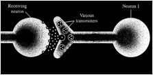 .
.
We
have a number of different kinds of neurotransmitters. Glutamate is an
excitatory neurotransmitter that is found throughout the nervous system. GABA
(gamma aminobutyric acid) is inhibitory and, like glutamate, is found
everywhere in the nervous system. Certain neurotransmitters are more specific
in what they communicate. For example, dopamine is related to pleasure and
reward. Serotonin involves emotionality and sleep patterns, norepinephrine
influences alertness, and endorphins alleviate pain. The neurotransmitter
system usually has everything it needs already built in. But when the
neurotransmitters are out of balance, drug therapy and psychological treatments
such as meditation, hypnosis, and psychotherapy can stimulate or inhibit
processes to stimulate a better balance. Medications for psychological problems
such as depression and anxiety are just acting to stimulate the neurons to
produce more of certain neurotransmitters or to block the action of other
neurotransmitters.
The
transmission across the synapse either activates the neuron to fire or
deactivates it from firing. When neurons fire together repeatedly, these
neurons tend to become wired together, known as Hebb’s Rule, forming a stronger
synaptic connection. This firing and wiring strengthening, known as LTP (long
term potentiation), explains, at a neuronal level, how learning and memory
occur. It also helps to account for neuroplasticity.
The
Central Nervous System and
Peripheral
Nervous System
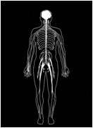
All the
neurons combined make up the nervous system, consisting of the central nervous
system (brain and spinal cord) and the peripheral nervous system (autonomic
nervous system, cranial nerves, and spinal nerves). The peripheral nervous
system extends through the whole body and communicates information to and from
the central nervous system. The autonomic nervous system interacts closely with
the central nervous system, often automatically and unconsciously.
The
neurons of the autonomic nervous system include two key systems: the
sympathetic nervous system and the parasympathetic nervous system. The
sympathetic nervous system prepares the body for vigorous action. The
parasympathetic nervous system acts as an opposite to the sympathetic nervous
system’s activations. So, when the sympathetic activation constricts blood
vessels or inhibits digestion during exercise, the parasympathetic system
relaxes vessel walls and stimulates digestion when the workout is over. Both
systems work together to help foster appropriate
responses. These systems of activation and deactivation are involved in
emotions such as fear and anger, as well as participating in responses to
stress and feelings of enjoyment. Together, these two systems
maintain the control that keeps the mind, brain, and body in balance. Yoga breathing, postures, and
meditations can shift the balance in the autonomic nervous system.
Brain
Structures and Functions
The
brain orchestrates the nervous system. It is often described in terms of its
structures and functions. Unconscious processing tends to travel a short,
subcortical path through the lower brain areas, known as bottom-up processing
that does not engage the higher level-processing cortex. For awareness of
emotions, sensations, and cognitions, the information usually travels a long
path, sometimes called top-down processing, involving higher parts of the
cortex.
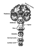
Lower
Brain Areas: Brainstem and Cerebellum
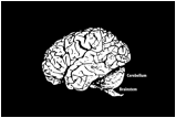
At the
base of the brain is the brainstem, the transition between the spinal cord and the
brain. This area is important in regulating vital body functions such as
breathing, heart rate and other automatic functions. These lower brain areas
coordinate their action with many other regions of the brain.
The
cerebellum (Latin for little brain), located at the back of the neck, has two hemispheres
with functional sections in each, known as lobes. The cerebellum interacts
closely with other parts of the brain through loops of interaction. It serves a
variety of functions including the regulation of higher cerebral processes in
motor planning, cognition, involuntary functions, and problem solving. It also
regulates posture and the command of movement. We have all experienced the
effort required to learn new movements, such as playing a sport or mastering a
dance pattern. During the learning period, the cerebellum is active. Once
movement control is mastered, the cerebellum becomes less active and other
parts of the brain get involved.
Interior
Brain Areas: Basal Ganglia and Limbic System
The
region that spans the area from the brainstem to below the cortex in the
interior of the brain is the limbic system for emotions and the basal ganglia
for voluntary movement and coordination.
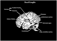
The
basal ganglia form a C-shape of four interconnected structures: the substantia
nigra, the caudate nucleus, the putamen, and the globus pallidus. These
structures are also involved in planning movement, performing movements in
sequence, and maintaining learning. This area is also part of predictive
control, attention, and working memory.
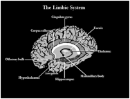
The
limbic system has been given much attention for therapy since it is intimately
involved in regulating emotion, fear conditioning, fight or flight and stress
responses as well as learning, and memory. Many structures play a central role in the limbic system.
The most central ones include the amygdala for emotions, the hippocampus for
learning and memory, the hypothalamus for regulating many autonomic functions
including biological rhythms and stress, and the thalamus as a gateway for
sensory information. Several other structures are considered important for some
aspects of emotion, and thus are the olfactory cortex, involved in the sense of
smell, the pituitary gland regulating hormones, and the nucleus accumbens,
important for reward, laughter, pleasure, addiction, and the placebo effect.
Two cortical areas are also strongly linked to the limbic system: the cingulate
gyrus for monitoring conflicts and the orbitofrontal cortex (part of the
pre-frontal cortex). All of these structures interconnect and interact
together, although some contribute more to one function than to another. With
so many varied brain structures all closely interacting functionally with each
other as well as with higher cortical functions, it makes sense as to why
emotions play such an important role in every aspect of living.
Higher
Brain Areas: The Cerebral Cortex
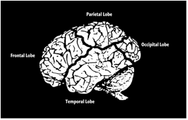
The
cerebral cortex is the outer layer of the hemispheres with many convolutions, gyri, and
folds, sulci. Folding increases the surface area of
the cortex, so that more than two thirds of the surface is hidden from view.
The cortex is sometimes referred to as the higher part of the brain. Each
hemisphere is divided into four lobes: the frontal, parietal, temporal, and
occipital. The lobes monitor different functions, although they are all
interrelated, interacting together.
The fibers that connect the two hemispheres are called the corpus
callosum, which helps the two hemispheres to be able to communicate with each
other.
Frontal
Lobe
The
frontal lobe accounts for nearly one-third of the cerebral cortex. The
prefrontal cortex is located at the front of the frontal lobe behind the
forehead. The fact that the prefrontal areas have extensive links throughout
the brain shows how interrelated brain functions really are. Areas of the
prefrontal cortex are involved in executive functions that include planning,
higher-level decision-making, sequencing, and goal directed behavior.
Independent thinking is processed in this area as well. Another part of the
prefrontal cortex processes personality characteristics such as the experience
of empathy, socially appropriate behavior, and emotional control.
The
primary motor area of the frontal cortex is located in the back (posterior)
area of the frontal lobe. It is
important for control of movement. This area has a map of the body on it.
Larger portions of the cortical map are devoted to areas that are used more,
such as the hands and face. The non-primary motor cortex, located in front of
the primary motor cortex, includes a pre-motor area and a supplementary motor
area. These areas are involved with movement and coordination in general, such
as stance, gait, and initiation of voluntary movement sequences. Mirror
neurons, are located in the motor area of the frontal lobe as well. These
neurons are involved in understanding and empathizing with the intentions and
actions of others and help with social understanding.
One
other important area located in the frontal lobe is the cingulate gyrus, also
called the cingulate cortex. This area is involved in motivated behavior,
spontaneity, and creativity. Complex behavior and attention or conflict
monitoring, are also processed in the cingulate gyrus. This area is primary
during the emotional reaction to pain and in the regulation of aggressive
behavior. It has also been found to play an important role in maternal
attachment as evident in behaviors like nursing and nest building in
animals.
The
Parietal Lobe
Behind
the frontal lobe and close to the cingulate gyrus is the parietal lobe. This
lobe is involved in sensation and perception of touch, pressure, temperature,
and pain. Sensory information from the body is correlated there during
perception or cognition of a sensation. The parietal lobe is activated when
locating objects in space and mapping the relationship of the body to the
world. The back (anterior) portion of the parietal lobe is the sensory strip.
The body is mapped on the sensory strip for sensations, similar to how the
primary motor cortex is where movement is mapped for the body.
The
Temporal Lobe
The
temporal lobe houses the primary auditory cortex. It is located near the
temples and moderates auditory information. Wernicke’s area, on the left
hemisphere side, plays a larger role in understanding spoken language. Although
most of the visual processing occurs in the primary visual areas in the
occipital lobe, some visual processing is performed in the temporal lobes,
involving perception of movements and face recognition.
The
Occipital Lobe
The
occipital lobes are located in the posterior region of the brain. Axons coming
from visual input from the eyes pass through the thalamus and are directed to
the primary visual cortex. The visual cortex is also sometimes called the striate cortex
because of its striped appearance. Human beings rely on their vision quite
heavily, and this is revealed in the complexity of this region of the brain.
There are more than thirty-two zones for visual processing differentiating
different aspects of seeing such as color, texture, and movement. All are located in the occipital lobes.
How
the Brain Areas Work Together
So,
how do all of these brain areas function? Senses provide a window to the world.
First, the receptor organs (such as the eyes, fingertips, nose, tongue or ears)
detect a stimulus. Each sensory system has its own pathway that sends the
signal to the cortex. The signal registers on receptor fields for the
particular sensory modality in a cortical map, located on the cortex. Maps can
change depending on what stimuli are experienced. Maps that are used more often
tend to grow larger. Attention, regulated in the frontal lobe, is what helps to
notice important stimuli and ignore others, such as attending to reading while
ignoring rain sounds on the roof.
Pathways
The
central nervous system is a complex collection of structures and functions that
are organized in pathways. Thoughts, feelings, and behaviors are intimately
involved in the flow of these pathways, dynamic systems of interactions between
brain structures and the flow of energy and neurotransmitters.
A
number of pathways through the nervous system help to coordinate the
mind-brain-body balance.
One
pathway processes sensory input and has a special pathway to process painful
stimuli.
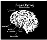
A reward
pathway regulates positive emotions and drives toward fulfillment,
satisfaction, and enjoyment.
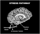
The
fear pathway, also called the HPA pathway, provides the capacity to respond to
threat and then return to homeostatic balance. (When over-activated, the fear
pathway becomes a stress pathway. Regulatory systems control the appetite and
the sleep-wake cycle as well. When any of these systems are out of balance,
disorders and problems tend to develop. All of these systems can be altered
using the therapeutic use of meditation, hypnosis, as well as psychotherapy.